TBCT: basic concept
Enabled by the recent development of X-ray flat panel imager (FPI) technology, cone-beam CT (CBCT) has quickly become the primary onboard imaging modality for image-guided radiotherapy (IGRT). CBCT has a compact geometry and can be readily integrated with radiation treatment machines. However, the image quality of CBCT is unsatisfactory due to the problems such as excessive X-ray scattering, suboptimal detector performance, etc. We have proposed to develop a novel TBCT system that is immune to the problems of the CBCT.
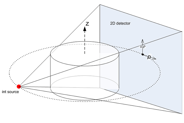
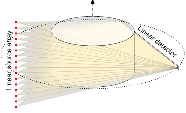
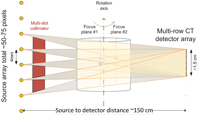
TBCT employs a linear array of discrete X-ray sources and a linear detector array. At one gantry angle, a sequential scan of all source pixels forms the shape of a tetrahedron in contrast to the cone of CBCT. TBCT uses the same high-quality detector array as fan-beam CT (FBCT). A multi-slot collimator collimates the X-ray beams to a stack of fan beams. Although a single-row detector is sufficient for 3D reconstruction, a multi-row detector can achieve isotropic image resolution in 2D and 3D imaging.
We previously developed a TBCT system with a low power multi-pixel thermionic X-ray (MPTEX) source. As shown in the picture, TBCT image quality is superior to the CBCT, but its resolution is still lower quality than the helical CT. TBCT image quality can be further improved with higher tube power and smaller detector pixel size. We are currently building a new TBCT benchtop system with the MPTEX source that we developed.
TBCT benchtop
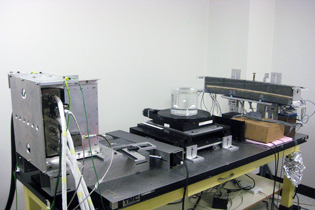
We developed a TBCT benchtop system using a MPFEX source (XinRay system) and in-house developed 5-row CT detector. Now we are developing another TBCT benchtop system with a newly developed high power MPTEX source.

Dual source TBCT for image guided radiotherapy (IGRT)
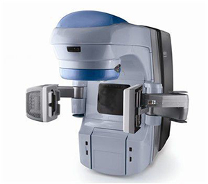
Besides inferior image quality, CBCT also cannot detect target motion along the kV imaging beam when mounted orthogonally to the MV treatment beam. We propose mounting the dual-source in-line, as shown in the picture. Not only can dual-source TBCT produce high-quality volumetric images, it can also produce stereoscopic images for real-time imaging during treatment delivery.
Collaborators
Mark Anastasio, PhD (Department of Biomedical Engineering)
David Politte, DSc (Mallinckrodt Institute of Radiology)
Hua Li, PhD (Department of Radiation Oncology)
Sasa Mutic, PhD (Department of Radiation Oncology)